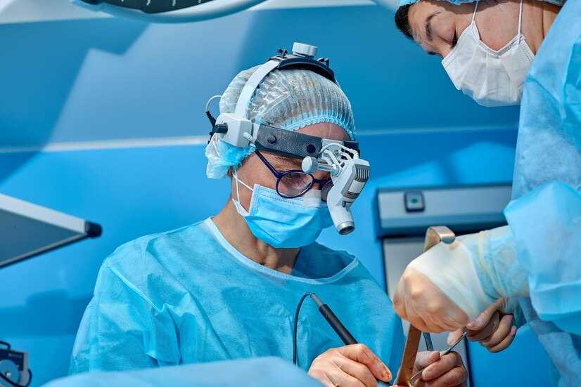
Surgery For Retinal Detachment

Introduction
Surgery for retinal detachment is a critical procedure aimed at restoring the attachment of the retina to the underlying tissues of the eye. Retinal detachment occurs when the retina separates from its normal position, leading to vision loss if left untreated. Surgery is necessary to prevent permanent vision impairment and preserve visual function.
Indications for Surgery
Symptomatic Retinal Detachment
Symptoms of retinal detachment include sudden onset of floaters, flashes of light, and a curtain-like shadow over the field of vision. Surgery is indicated when these symptoms occur, indicating a high risk of vision loss.
High-Risk Factors
Certain factors increase the risk of retinal detachment, such as a history of trauma to the eye, severe myopia, previous eye surgeries, or family history of retinal detachment. Patients with these risk factors may require prophylactic surgery to prevent detachment.
Surgical Techniques
Scleral Buckling
Scleral buckling involves placing a silicone band (buckle) around the circumference of the eye to indent the sclera and relieve traction on the detached retina. This technique helps close retinal breaks and reattach the retina to the underlying tissue.
Vitrectomy
Vitrectomy is a surgical procedure that involves removing the vitreous gel from the eye and replacing it with a gas bubble or silicone oil. This allows the surgeon to access the retina and repair retinal tears or detachments using laser photocoagulation or cryotherapy.
Pneumatic Retinopexy
Pneumatic retinopexy is a minimally invasive procedure used for select cases of retinal detachment. It involves injecting a gas bubble into the vitreous cavity to push the detached retina against the eye wall, followed by laser or cryotherapy to seal retinal breaks.
Postoperative Care
Positioning
After surgery, patients may need to maintain a specific head position to ensure the gas bubble or silicone oil tamponade holds the retina in place. This positioning is crucial for successful reattachment of the retina.
Follow-Up Visits
Regular follow-up visits are essential to monitor healing and assess visual recovery. Patients undergo comprehensive eye examinations to evaluate the status of the retina and intraocular pressure.
Complications and Risks
Proliferative Vitreoretinopathy (PVR)
PVR is a common complication of retinal detachment surgery, characterized by the formation of scar tissue on the retina. This can lead to recurrent detachment and vision loss if not adequately managed.
Infection and Inflammation
As with any surgery, there is a risk of infection and inflammation following retinal detachment surgery. Patients are prescribed antibiotics and anti-inflammatory medications to reduce the risk of complications.

